4D Endoscopic Ultrasonography
We are developing a pseudo-3D visualization system for endoscopic ultrasonography by superimposing ultrasound images. The system comprises an ultrasonic endoscope and an electromagnetic tracking sensor. A pseudo-3D volume is reconstructed by integrating ultrasound images captured at arbitrary positions by the endoscope, and the resulting volume is overlaid onto endoscopic images in real time for the surgeon.
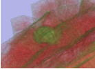 3D model of gallbladder |
4D Endoscope |
Papers
- Yumi Iwashita, Shinji Tarumi, Ryo Kurazume, Makoto Hashizume, Development of Pseudo 3D Visualization System by Superimposing Ultrasound Images, International Symposium on Assistive and Recuperative Technologies for Injured, Ill, Pregnant, Elderly and people with Disabilities, Isai, Romania, July 24-26, 2009
- Shinji Tarumi, Yumi Iwashita, Ryo Kurazume, and Makoto Hashizume, Development of Pseudo 3D Visualization System for Endoscopic Ultrasonography, Proc. 5th Joint Workshop on Machine Perception and Robotics (MPR2009), MPR2009-ps2-5, (2009 10)
Patient-specific 3D bone shape estimation of femur from two radiographs
We are conducting joint research with the Medical School of Kyushu University and Osaka University. Research topics include a navigation system for endoscopic robotic surgery, a 3D diagnostic system for hip replacement surgery, an image-guided system for endotracheal intubation, and more. As an example, we propose a method for estimating the 3D shape of a patient's femur using two radiographs and a parametric femoral model. First, the parametric model is constructed through statistical analysis of 3D femoral models derived from CT scans of multiple patients. Then, both the pose and patient-specific shape parameters of the model are estimated from the two 2D radiographs using a distance map generated by the Level Set Method. We have conducted experiments using synthesized images, fluoroscopic images of a femur phantom, and in vivo images from hip prosthesis patients. The results demonstrate that the proposed system is practically applicable and effective for clinical use.
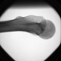 Radiograph |
Shape estimation |
Papers
- Ryo Kurazume, Kaori Nakamura, Toshiyuki Okada, Yoshinobu Sato, Nobuhiko Sugano, Tsuyoshi Koyama, Yumi Iwashita, Tsutomu Hasegawa, 3D reconstruction of a femoral shape using a parametric model and two 2D fluoroscopic images, Computer Vision and Image Understanding, Volume 113, Issue 2, pp. 202-211, (2009.2)
- Yumi Iwashita, Ryo Kurazume, Kaori Nakamura, Toshiyuki Okada, Yoshinobu Sato, Nobuhiko Sugano, Tsuyoshi Koyama , Tsutomu Hasegawa, Patient-specific femoral shape estimation using a parametric model and two 2D fluoroscopic images, ACCV'07 Workshop on Multi-dimensional and Multi-view Image Processing, MM-O-9, November 2007.
- Ryo Kurazume, Kaori Nakamura,Toshiyuki Okada, Yoshinobu Sato, Nobuhiko Sugano,Tsuyoshi Koyama, Yumi Iwashita, and Tsutomu Hasegawa, 3D reconstruction of a femoral shape using a parametric model and two 2D fluoroscopic images, in Proc. IEEE International Conference on Robotics and Automation, 2007.
- Yumi Iwashita, Shinji Tarumi, Ryo Kurazume, Makoto Hashizume, Development of Pseudo 3D Visualization System by Superimposing Ultrasound Images, International Symposium on Assistive and Recuperative Technologies for Injured, Ill, Pregnant, Elderly and people with Disabilities, Isai, Romania, July 24-26, 2009
Surgical robot navigation
In collaboration with Professor Makoto Hashizume of the Kyushu University Faculty of Medicine, Professor Motoji Yamamoto of the Faculty of Engineering at Kyushu University, and Professor Kazuo Kiguchi of the Graduate School of Science and Engineering at Saga University (currently Kyushu Unversity), we are developing a surgical navigation system for the Da Vinci surgical robot.
Papers
- Ken'ichi Morooka, Xian Chen, Ryo Kurazume, Uchida Seiichi, Kenji Hara, Yumi Iwashita, Makoto Hashizume, Real-time Nonlinear FEM with Neural Network for Simulating Soft Organ Model Deformation, The 11th International Conference on Medical Image Computing and Computer Assisted Intervention (MICCAI 2008), New York, USA, Sep. 2008
2D-3D Registration for Navigation System of Surgical Robot
We have proposed a novel registration algorithm for aligning 2D color images with 3D geometric models for use in surgical robot navigation systems. The 2D–3D registration process is essential for accurately superimposing a tumor model onto endoscopic images, and thus plays a critical role in the effectiveness of surgical navigation. Consequently, the performance of the registration algorithm directly impacts the usability of the surgical robotic system. A commonly used technique involves external markers; however, its accuracy is often compromised by patient movement due to respiration, heartbeat, or other unpredictable factors. To achieve precise registration without relying on external markers or specialized measurement devices, we propose a new method that leverages 2D images and their corresponding distance maps, which are generated using the Fast Marching Method.
| 2D-3D registration | Tracking 3D object |
| Endoscopic image | Endoscopic image |
Papers
- Yumi Iwashita, Ryo Kurazume, Kozo Konishi, Masahiko Nakamoto, Naoki Aburaya, Yoshinobu Sato, Makoto Hashizume and Tsutomu Hasegawa, Fast Model-Image Registration using 2D Distance Map for Surgical Navigation System, Advanced Robotics, Vol.21, No.7, pp751-770, (2007.4)
- Yumi Iwashita, Ryo Kurazume, Kozo Konishi, Masahiko Nakamoto, Makoto Hashizume, and Tsutomu Hasegawa, Fast 2D-3D Registration for Navigation System of Surgical Robot, in Proc. IEEE International Conference on Robotics and Automation, pp.909-915, 2005.
- Yumi Iwashita, Ryo Kurazume, Kenji Hara, and Tsutomu Hasegawa, Fast Alignment of 3D Geometrical Models and 2D Color Images using 2D Distance Maps, in Proc. The 5th International Conference on 3-D Digital Imaging and Modeling, 2005.