Morooka Lab: Image information and robotics, Kyushu University

MEDICAL IMAGE PROCESSING
In medical image processing group, we are developing a diagnosis and theraphy support system using image information technology such as image processing, computer vision, computer graphics, virtual reality, mixed reality, and so on.
- Real-Time Nonlinear FEM (Finite Element Method)-based Simulation of Soft Tissue Deformation
- Digital Brain Atlas of Japanese People for Safe and Accurate Stereotactic Neurosurgery
Real-Time Nonlinear FEM (Finite Element Method)-based Simulation of Soft Tissue Deformation
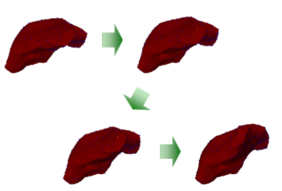
In the field of 3D object modeling, the modeling of soft changed tissue changing as time goes on is one of hot research topic.
There are two (3D) modeling methods for physical actions which are Nonlinear Finite Element Method(FEM) and high precision dynamic simulation.
Despite of satisfactory results on both methods, those require a large amount of computing resources.
Recently, the speed up of FEM is available using approximate calculation, but it still has a problem to decrease the estimation accuracy.
To solve that problem, we implemented a neural network based 3D modeling system which has similar performance with nonlinear FEM and also guarantees real-time processing.
From that system, now we are developing a new low-invasion operation simulator.
See the detail of the research ->> Click
Digital Brain Atlas of Japanese People for Safe and Accurate Stereotactic Neurosurgery
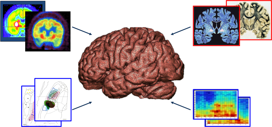
Deep Brain Stimulation(DBS) is a surgical treatment involving the implantation of a medical device called a brain pacemaker,
which sends electrical impulses to specific parts of the brain. DBS in select brain regions has provided remarkable thera- peutic benefits
for otherwise treatment-resistant movement and affective disorders such as chronic pain, Parkinson's disease, tremor and dystonia.
Practically, a classical brain atlas has been used by altering three dimensional magnification to fit patients’ brain images.
However, it is difficult to apply the classical atlases to the individual Japanese patient because coronal,
horizontal and sagittal slices of the atlases are made from different Westerners.
Together with our collaborators at the Faculty of Medicine, Kyushu University and Kumamoto University, we
are developing the Digital Brain Atlas for Japanese combined with shape, dissection, and functional data.
See the detail of the research ->> Click